Magnetic Resonance Imaging (MRI)
is a modern examination method that allows to diagnose pathological changes in human bodies. During the examination, images of the internal organs are produced, which help to reveal the cause of the condition, establish a diagnosis and initiate timely and appropriate treatment. The examination does not use X-rays but a strong magnetic field. The MRI examination method is therefore free of any harmful effects.
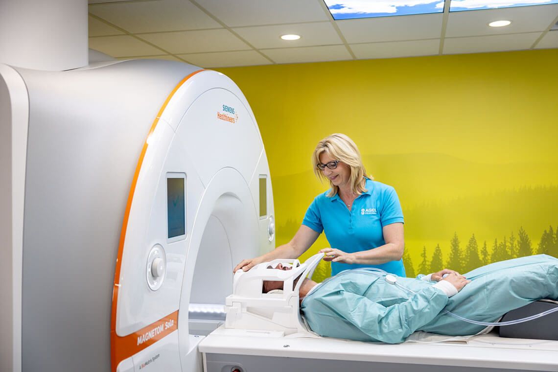
At our workplace, we perform examinations using a modern machine – Magnetom Sola 1.5T from Siemens.
Advantages of the examination:
- Magnetic resonance imaging is a fast and non-invasive method.
- The imaging of soft tissues such as brain and spinal cord tissue, muscles, tendons, joints and others is much more detailed and pronounced than with other methods.
- Contrast agents (paramagnetic) cause far fewer allergic reactions than the iodine contrast agents used in conventional X-ray and CT scans.
- The image can easily be made in all 3 basic planes.
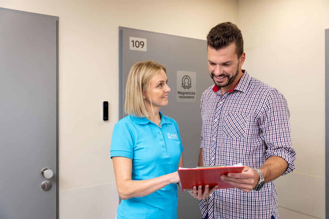
What to bring for the examination
Bring a valid health insurance card, a properly completed request form for the examination from the referring physician, which correctly states your personal details, specifies the examination required and, where appropriate, the expected benefit. It is always necessary to mention any surgeries and procedures that may have left metallic material in the patient's body.
If you have previously been examined at another healthcare facility, it is advisable to bring the relevant image documentation on a CD with you or ask the site of the previous examination to send it electronically via ePACS network.
How to prepare for an MRI scan
- No special preparation is usually necessary before an MRI scan.
- It is advisable not to overeat, as you will be lying motionless on the examination table for an extended period of time – approximately 20 to 50 minutes.
- An exception is an abdominal MRI scan – e.g., kidney, liver, pancreas, intestines and biliary tract (MRCP) – where you should not drink or eat for at least 3-4 hours before the examination. The permanent medication may be taken and washed down with water. When it comes to the taking images of the small intestine (MR Enterography), it is necessary to arrive 90 minutes before the examination itself.
- For all other examinations, you only need to arrive 15 minutes in advance.
- We recommend not to use make-up products before the examination, as some may contain iron compounds and thus distort the results. Clothing should not contain metal buckles, hooks, or decorative threads, even on underwear. This also applies to underwired bras, so we recommend wearing sports bras without underwires for the examination.
- Use the toilet just before the examination. For the pelvic imaging, the bladder should be moderately full, and you should urinate about one hour before the examination.
- Before you lay down on the examination table, you will remove some of your clothing. You will keep your underwear, T-shirt, and socks (if these items of clothing do not have metal components), otherwise you will remove them too and receive an examination gown, and you will be instructed by the MRI staff on how to behave during the examination.
- All metal objects cause significant distortion of the results. If there is an uncertainty, you may be sent for a follow-up X-ray to clarify whether it is a metallic object and where it is located.
Each patient must have a written informed consent completed and signed before undergoing an MRI scan.
Magnetic Resonance Imaging – the course of examination
When examining minors, the consent of the legal guardian is required. We examine cooperating children from 5 years of age. The parent (legal guardian) may be present during the examination of his/her child.
You will be placed on an examination table by a radiology assistant who will move the table into the opening of the machine. Most examinations are performed with the patient lying on their back, in some cases lying on their stomach. The examination itself is painless, but you might feel warmth during it. It is necessary to lie motionless the entire time and follow the instructions of the staff, e.g., to hold your breath, breathe in or out when prompted. The staff will thoroughly explain everything to you, and you may ask anything again just before the examination.
You will be fitted with noise reducing headphones, through which you can also communicate with the staff. You will also receive a signal balloon in your hand, and you can press it to call the operator in case of any problem.
In most cases, the MRI is performed without a contrast agent applied intravenously, but in some examinations it is added as part of the ongoing scan to clarify the findings. It is well tolerated by patients; allergic reactions are very rare.
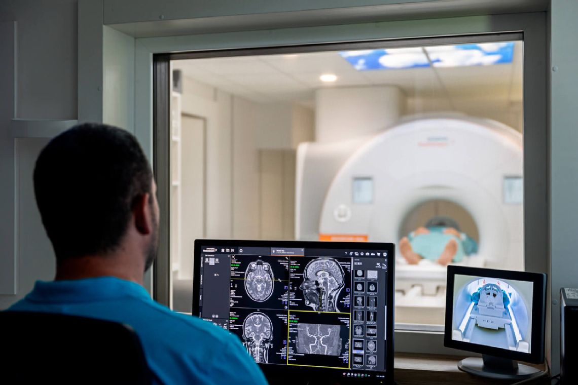
Result of the examination
The imaging part – immediately after the examination:
- At the request of the referring physician, via e-PACS
- Uploaded to a flash drive purchased here (according to the current price list) and handed over to the patient
The result in written form is produced within 7 working days.
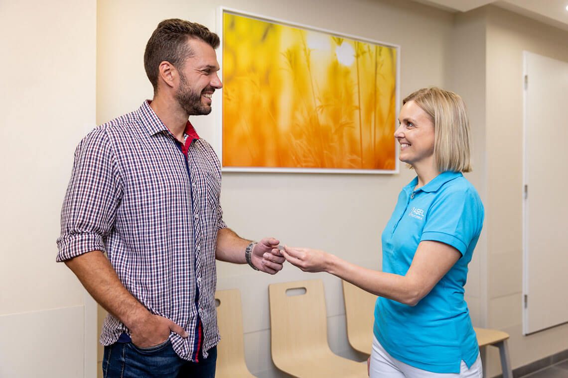
Delivery:
- To the referring physician by mail.
- Personal collection by the patient at the reception of the polyclinic against the presented ID card. If you are unable to collect your finding in person, you must give your power of attorney to the authorised person.


Indication (reason)
Tumors, inflammations, post-traumatic changes, MS, vascular diseases and others.
- Eye sockets (orbits)
- Paranasal sinuses (VDN)
- Hypophysis
- MR angiography of intracranial arteries
- MR venography
Thyroid gland, nodules, resistance, and other.
Degenerative disorders, inflammation, tumours, post-traumatic changes.
- Cervical (C) spine
- Thoracic (Th) spine
- Lumbosacral (LS) spine
- Coccyx and sacrum
Degenerative changes, traumatic changes (of bones, cartilage, ligaments, muscles), inflammation, tumours.
- Hip joints
- Sacroiliac joint (SIJ)
- Knee joint
- Shoulder joint
- Other joints – elbow, wrist, hand, ankle, Achilles tendon, foot-instep, fingers and toes
- Long bones – arm, forearm, thigh, lower leg
- Kidneys
- Liver
- Intestines – enterography
- Pancreas
- Spleen
- Biliary tract – MRCP
Degenerative and post-traumatic conditions, cancer and inflammatory diseases, examination of nerves, muscles, muscle attachments.
- Anus – rectum
- Fistula
- Pelvis minor
- Clear bony pelvis
- Prostate gland and others
Contraindications (circumstances precluding the examination)
Pacemaker, cochlear implant, neural stimulator, insulin pump, and metal chips in the eye. You must not be examined with these items in your body.
Newly introduced metals, clamps, or prostheses. We perform the examination with an interval of 6 weeks from the implantation.
In the first trimester, MRI is not performed at all; if necessary, MRI can be performed at a later stage of pregnancy, but only natively (without the use of a contrast agent). The application of gadolinium contrast agent is absolutely contraindicated; the substance passes through the placenta into the foetus.
The gadolinium contrast agent administered intravenously passes into breast milk. It is recommended to express breast milk before the examination and not to breastfeed for 24 hours after the examination, but to express milk to cleanse the mammary glands. Native MRI can be performed without limitations.
The patient can be prepared for the examination in advance using corticosteroids.
The application of a contrast agent may only be performed in justified cases. Adequate hydration is necessary.
They must inform their referring physician, who may administer anti-anxiety medication. However, in this case you should be accompanied and should not drive.
In case the patient does not fit into the tunnel of the MRI machine. The diameter of the tunnel – gantry – of our machine is 70 cm.
Foreign or implanted metal materials can cause damage to the MRI machine and the patient's health.
We do not perform examinations under general anaesthesia.
Furthermore, we do not perform examinations even with an MRI compatible pacemaker/implanted defibrillator, as it is necessary to switch it off or reprogram it in cooperation with the cardiology department.